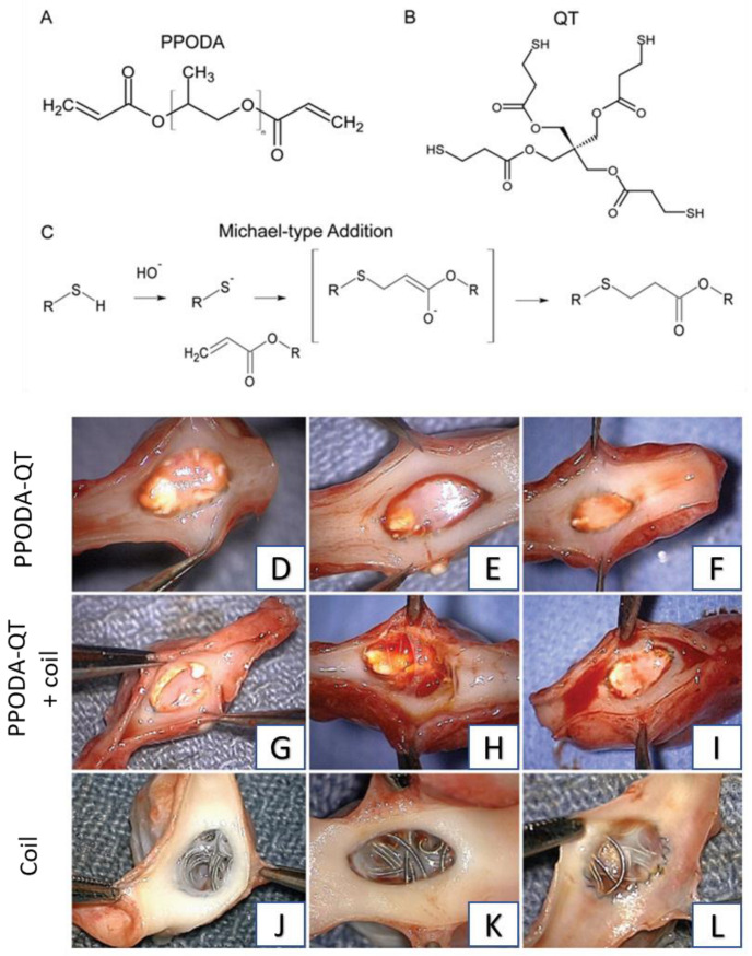
Figure 2.
Components (A,B) and reaction scheme (C) of PPODA−QT. Photographs of explanted aneurysm samples. The PPODA−QT aneurysm samples (D–F) all show a smooth surface in the ostium. The coil + PPODA−QT aneurysms (G–I) show excess PPODA−QT protruding into the parent vessel in two samples, resulting in rough surfaces (G,H), while one sample displays no PPODA−QT protrusion and a smooth surface (I). The coil-only aneurysms (J–L) show the least neointimal tissue overgrowth. Reprinted with permission from Ref. [86]. 2013, J. Neurosurgery.

