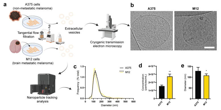
Figure 1.
Physicochemical characterization of extracellular vesicles (EVs) from human A375 melanoma cells and human M12 melanoma brain metastases cells. (a) Schematic representation of the origin and purification of A375 and M12 cell lines. (b) Cryogenic transmission electron microscopy images. Scale bar, 100 nm. (c) Size distribution profiles of EVs. (d) Concentration of EVs. (e) Average modal diameter of each sample. Data are presented as the mean (unless otherwise noted) + SD of five replicates. Statistics are based on a non-parametric Mann-Whitney test. *, p < 0.05; **, p < 0.01; ****, p < 0.0001.

