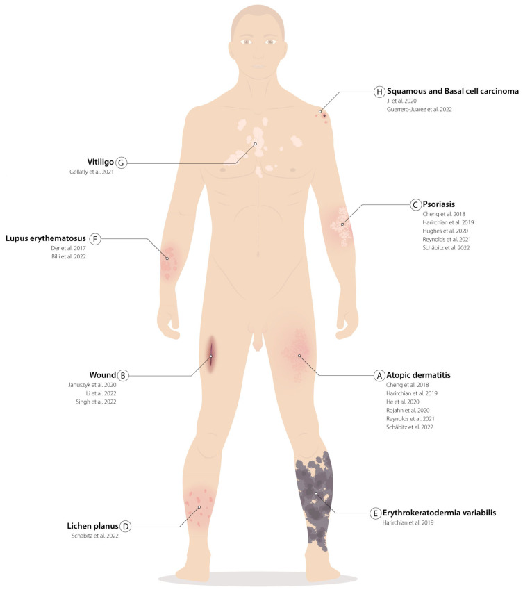
Figure 2.
Skin pathological tissues analyzed in single cells: (A–H) List of skin diseases that have been characterized with single-cell techniques. For each disease, references to the papers that have characterized the specimens are listed in chronological order. Localization of the illustrated pathologies does not reflect the actual position from which the biopsies were withdrawn but serves as an example. (A) Atopic dermatitis [20,25,30,31,46,47]; (B) Wound [37,41,50]; (C) Psoriasis [25,31,46,47,51]; (D) Lichen planus [46]; (E) Erythrokeratodermis variabilis [47]; (F) Lupus erythematosus [42,48]; (G) Vitiligo [29]; (H) Squamous and basal cell carcinoma [34,49].

