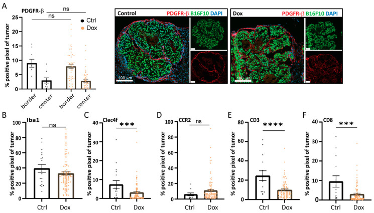
Figure 5.
Increased TGF-β receptor-I signaling in B16F10 alters the tumor microenvironment in liver metastases. Analysis of the immunohistochemical staining of microenvironmental cell markers in B16F10 caALK5 liver metastases treated with and without dox. All metastases per group are shown, from n = 9–14 in the control and n = 84–101 in the dox-treated group. The percentage of positive staining pixels in metastases was calculated; analysis was performed using QuPath. Mean ± S.E.M. Significance was calculated using Student’s t-test, ns, *** p ≤ 0.001, **** p ≤ 0.0001. Staining was performed for the CAF marker PDGFR-β (A), general macrophage marker Iba1 (B), liver-resident macrophage marker Clec4f (C), bone marrow-derived macrophage marker CCR2 (D), general T cell marker CD3 (E), and cytotoxic T cell marker CD8 (F).

