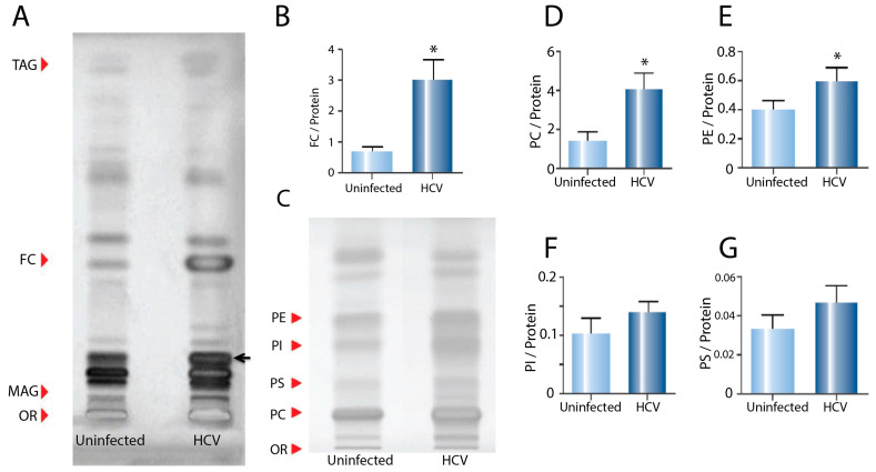
Figure 2.
Effect of HCV infection on neutral lipid and phospholipid species in endoplasmic reticulum fractions. (A) HPTLC plate loaded with neutral lipids from ER extracted from uninfected control and HCV-infected cells (OR—origin; MAG—monoacylglycerol; FC—free cholesterol; TAG—triacylglycerol). (B) Quantification of FC in the ER of uninfected control and HCV-infected cells. (C) HPTLC plate loaded with phospholipid species from ER extracted from uninfected control and HCV-infected cells (OR—origin; PC—phosphatidyl choline; PS—phosphatidyl serine; PI—phosphatidyl inositol; PE—phosphatidyl ethanolamine). (D) Quantification of PC, (E) PE, (F) PI and (G) PS. Analysis was performed using triplicate samples from three biological replicates, normalised for protein concentration (* p < 0.05).

