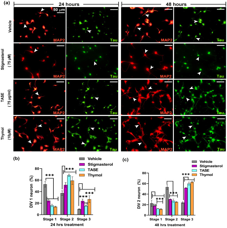
Figure 5.
Effects of TASE and thymol on early neuronal differentiation. Hippocampal cultures were incubated with TASE and thymol for 24 h and 48 h with vehicle and stigmasterol (75 µM) as control, with dendrite stained with MAP2 (red) as a dendrite-specific marker and tau (green) as an axon marker. (a) Immunofluorescence images showing neuronal differentiation and outgrowth (axonal and dendritic sprouting, indicated with arrowheads). Arrowheads indicate the axonal and dendritic processes of the neuron. Scale bar, 50 μm applies to all images. (b) Percentage of neurons that reached different developmental stages at 24 h and (c) 48 h of incubation. Statistical significance is compared to vehicle with the p-values: *** p < 0.001, and ns (not significant) (one- and two-way ANOVAs with Dunnett’s and Tukey’s multiple comparisons tests). Data points are represented as mean ± SD (n = 500 individual neurons from three independent experiments). SD, standard deviation.

