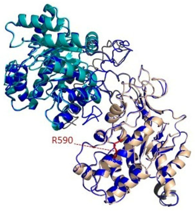
Figure 4.
Homology modelling prediction of MTHFR-L590R. Graphic representation of secondary structure of MTHFR mutated protein, modelled with I-tasser using X-ray structure of human MTHFR as template (6FCX PDB structure). The structure is composed by catalytic domain (represented in cyan), regulatory domain (represented in wheat) and the linker between them (represented in gray). The position of the arginine replacing the wild-type amino acid in patient is shown in red. The models of MTHFR have been prepared with PyMOL.

