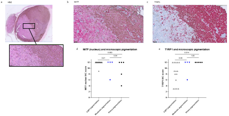
Figure 2.
Less MITF IHC staining is seen in heavily pigmented UM. (a): A UM case with two components with different pigmentation levels (haematoxylin and eosin) shows higher MITF staining in the light area than in the dark area of the tumour (b) and an opposite pattern in TYRP1 staining (c). (d,e): UM with more pigmentation show a lower IHC nuclear MITF score (d) and a higher TYRP1 score (e). Magnification: 1× is (a), 20× in (a) (detail, in black box), (b,c). H&E = Haematoxylin and eosin.

