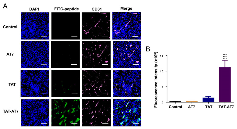
Figure 9.
TAT-AT7 targeted glioma blood vessels in the intracranial glioma tissue of nude mice. FITC-labeled AT7, TAT, and TAT-AT7 were injected into intracranial U87-mCherry-luc glioma-bearing mice via the tail vein. After 1 h, the glioma tissues of nude mice were taken for frozen sectioning. (A) The fluorescence distribution of tissues was observed under a panoramic scanning microscope after CD31 immunofluorescence staining. Blue fluorescence: nucleus stained with DAPI, green fluorescence: FITC-labeled peptide, pink fluorescence: CD31 stained with Cy5.5. Bars represent 200 μm. (B) Statistical analysis results of FITC fluorescence intensity. Each data point represents the mean ± standard deviation. n = 3, *** p < 0.001 vs. control group; ### p < 0.05 vs. AT7 group; &&& p < 0.05 vs. TAT group.

