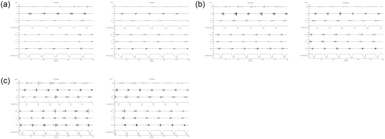
Fig 2. A representative electromyographic waveform in each muscle and strain gauge signals in the leading and trailing limbs.
(a) 7, (b) 10, and (c) 13 m/s canters. Br; Musculus brachiocephalicus, Inf; M. infraspinatus, TB; long head of M. triceps brachii, GM; M. gluteus medius, ST; M. semitendinosus, FDL; M. flexor digitorum longus. Forelimb strain and hindlimb strain refer to signals from stain gauges attached to the hooves.

