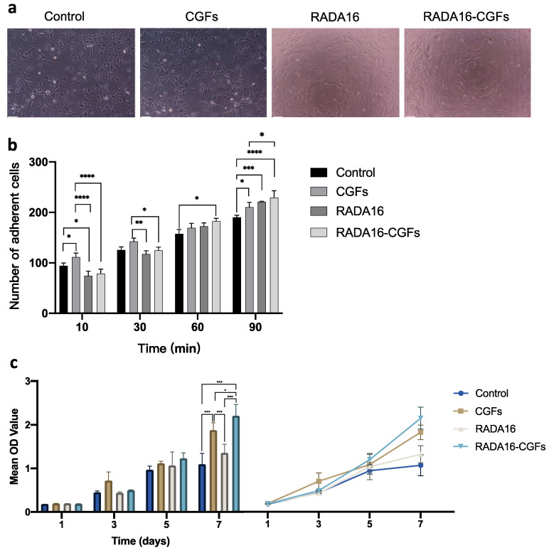
Figure 5.
Cell proliferation of MC3T3 with RADA16-CGF. (a) MC3T3 cell morphology co-cultured with different composites after 3 days. Scale bars, 100 μm. (b) Comparison of the number of adherent cells between the control, CGFs, RADA16, and RADA16-CGF groups at different timepoints. *, p < 0.05; **, p < 0.005; ***, p < 0.001; ****, p < 0.0001. (c) Comparison and tendency chart of the CCK-8 assays for the proliferation of MC3T3 cells co-cultured with different composites. *, p < 0.05; ***, p < 0.001.

