Figure 4.
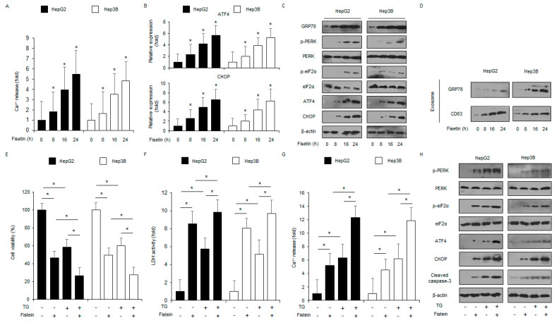
Fisetin induces ER stress in liver cancer cells. (A) HepG2 and Hep3B cells were treated with fisetin (100 μM) for the indicated times, and an intracellular Ca2+ assay was performed. * = p < 0.05. (B) The ER stress markers ATF4 and CHOP were measured by RT-PCR. Fold changes to target genes were normalized to β-actin. (C) The ER stress markers CHOP, PERK, eIF2α, p-eIF2α, p-PERK, ATF4, and GRP78 were measured by a western blot assay. β-actin was used as the protein loading control. (D) HepG2 and Hep3B cells were treated with fisetin (100 μM) for the indicated times, and then the exosomes (30 μg) were collected from the cell supernatant. Total exosomes were determined by western blotting using the exosome marker CD63 and the ER stress marker GRP78. (E–H) Cell viability, LDH activity, caspase-3 activity, intracellular Ca2+, and ER stress-related protein (cleaved caspase-3, p-eIF2α, ATF4, PERK, CHOP, eIF2α, and p-PERK) levels were measured in the thapsigargin (TG; 3 μM, 24 h) and fisetin (100 μM, 24 h)-treated HepG2 and Hep3B cells. * = p < 0.05. Data are representative of three experiments.

