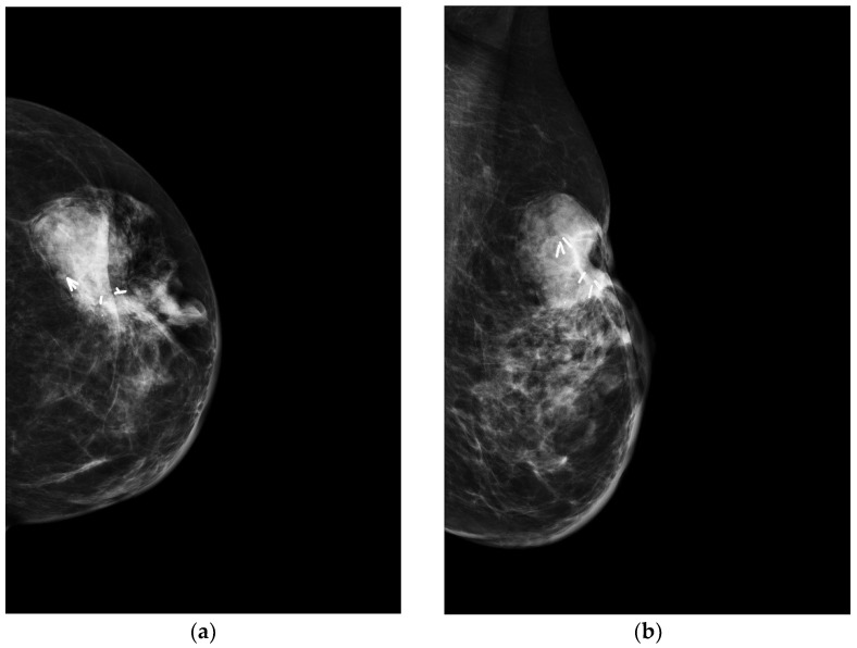
Figure 3.
A 70-year-old patient with a history of carcinoma of the left breast treated with quadrantectomy and radiotherapy. Three years after completion of radiotherapy, clinical examination revealed a reddish cutaneous area and a palpable mass in the surgical scar location. (a) Craniocaudal and (b) mediolateral oblique mammograms of the left breast show a high density, irregular mass below the scar associated to skin thickening. The white lines represent the surgical clips released at the time of the quadrantectomy.

