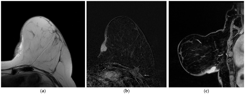
Figure 11.
A 48-year-old patient with a history of left breast cancer treated with quadrantectomy and radiotherapy. (a) T2-weighted, (b) axial 3D gradient echo T1-weighted post-contrast and (c) sagittal 3D gradient echo T1-weighted post-contrast images show skin thickening and an oval-shaped mass, with circumscribed margins, characterized by homogenous enhancement. US-guided core needle biopsy reveals SBA.

