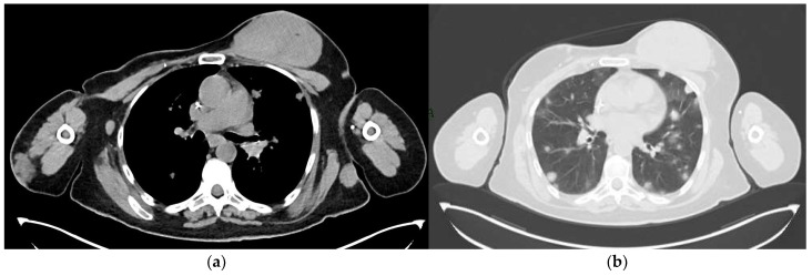
Figure 12.
A 65-year-old patient with a history of PBA treated with right mastectomy, right axillary lymph node dissection and radiotherapy of the right axilla. A 2-year follow-up with CT scan revealed metastatic disease: (a) Mediastinal window showing a voluminous mass in the left breast, an enlarged lymph node in the right axilla, and a nodular thickening in the subcutaneous soft tissues of the right arm and of the left lateral chest wall. (b) Parenchymal window showing numerous bilateral pulmonary nodules.

