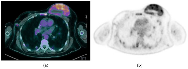
Figure 14.
A 65-year-old patient with a history of PBA treated with right mastectomy, right axillary lymph node dissection, and radiotherapy of the right axilla. A 2-year follow-up with 18F FDG PET-CT scan revealed metastatic disease: (a,b) hypermetabolic lesions are observed in breasts, lungs, and bones.

