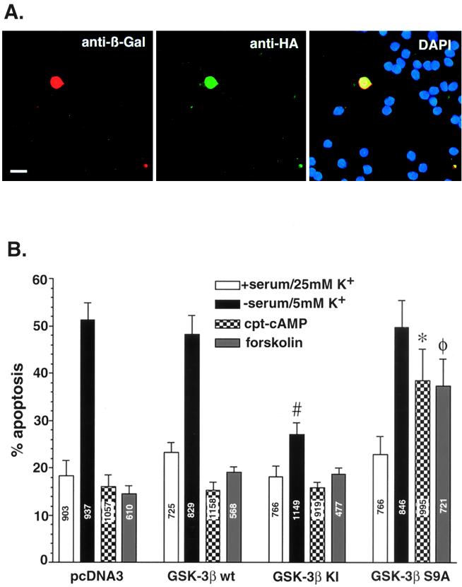
FIG. 6.
Transfection of cerebellar granule neurons with wt-GSK-3β, a KI GSK-3β mutant, and a Ser9→Ala9 GSK-3β mutant. Neurons were cotransfected with the control vector, wt GSK-3β, (KI) GSK-3β, (S9A) GSK-3β along with pCMV-β-Gal. One day after transfection, the neurons were placed in complete medium (serum, 25 mM KCl) or switched to serum-free medium containing 5 mM KCl, with or without cAMP (500 μM) or forskolin (10 μM). After 24 h the transfected neurons were fixed and immunostained with an antibody to β-Gal (Cy3-coupled secondary antibody) and 12CA5 antibody to HA (fluorescein isothiocyanate-coupled secondary antibody). To reveal nuclear morphology, neurons were also stained with DAPI. (A) Demonstration of the triple-staining method in neurons grown in serum and 25 mM KCl. Bar, 10 μm. (B) Effects of GSK-3β constructs on neuronal survival. The β-Gal-positive neurons were scored as healthy or apoptotic as described for Fig. 3C. Data are presented as means ± SEMs (n = 4). #, P < 0.001 versus pcFNA3 in pcDNA3 in apoptotic (serum-free, 5 mM KCl) medium; ∗, P < 0.001 versus pcDNA3 in apoptotic medium plus CPT-cAMP; φ, P < 0.001 versus pcDNA3 in apoptotic medium plus forskolin (Student's t test). Numbers on the bars indicate the total number of neurons counted.

