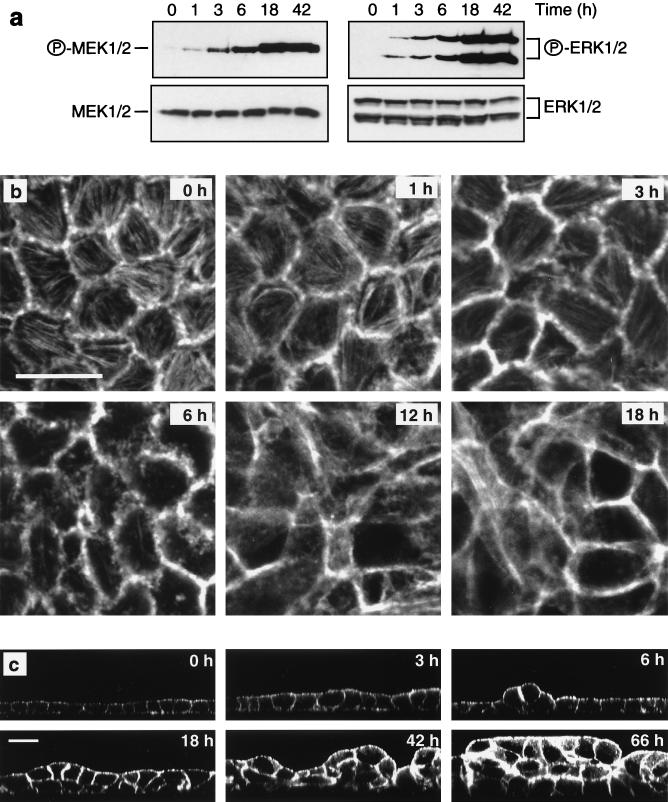
FIG. 1.
Time course of Raf-induced transformation of MDCK cells. Polarized MDCK cells expressing EGFPΔRaf-1:ER were treated with 1 μM 4-HT for the indicated periods of time. (a) Activation of MEK and ERK following Raf activation was assayed by Western blotting of whole cell lysates using activation-specific antibodies as described in Materials and Methods. (b and c) Confocal microscopy of MDCK cells labeled with fluorescent phalloidin revealed that the morphological alterations in response to Raf activation comprised progressive changes in cell shape (b and c), loss of actin stress fibers (b), increased cortical actin (b and c), and multilayering (c). (b) X-Y sections; scale bar, 20 μm. (c) X-Z sections; scale bar, 20 μm. All X-Y sections were sampled at the base of the cell monolayers.

