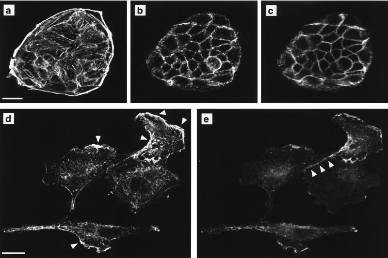
FIG. 6.
Raf transformation leads to spreading and scattering of MDCK cells. MDCK cells expressing EGFPΔRaf-1:ER grown at low cell density were either untreated (a to c) or treated with 1 μM 4-HT for 42 h (d and e). The cells were then fixed and labeled with fluorescent phalloidin to detect polymerized actin and immunolabeled to localize E-cadherin. (a and b) Phalloidin staining in optical sections through the basal (a) and apical (b) regions of untreated cells. (c) E-cadherin localization in the apical region of untreated cells. No E-cadherin labeling is detected in at the base of untreated cells (not shown). (d and e) Following activation of Raf, subconfluent MDCK cells become flattened, and both phalloidin (d) and E-cadherin (e) labeling is evident in a single optical section. Arrows in panel d indicate areas of apparent membrane ruffling. Note that E-cadherin colocalizes with polymerized actin at these areas. Arrowheads in panel e illustrate that E-cadherin is retained at areas of cell-cell contact in subconfluent Raf-transformed MDCK cells. Scale bars, 20 μm.

