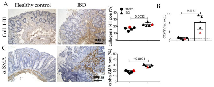
Figure 2.
Evaluation of pro-fibrotic signaling (collagens I-III, CCN2, α-SMA), in the colonic mucosa of IBD patients. (A) Immunohistochemistry and semi-quantitative analyses for collagens I-III (brown). (B) Quantitative Real-Time PCR for Cellular Communication Network Factor 2 (CCN2) expression. (C) Immunohistochemistry and semi-quantitative analyses for α-SMA. Nuclei are counterstained with hematoxylin; Original magnification 10×, scale bar 100 μm. These images are representative of at least n = 4 subjects/groups. Data were analyzed by unpaired t-test. IBD: inflammatory bowel diseases group. Red symbols indicated biopsies from healthy (red circle) and affected (red triangle) colonic mucosa of the same subjects.

