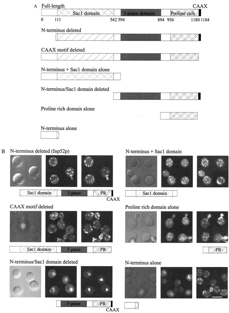
FIG. 6.
Analysis of Inp52p domains which mediate the intracellular localization in hyperosmotically stressed cells. Inp52p-GFP deletion mutant constructs were generated as described in Materials and Methods. (A) Schematic representation of wild-type and mutant Inp52p-GFP fusion proteins. 5-ptase, 5-phosphatase. (B) Confocal microscopy of yeast expressing mutant Inp52p-GFP fusion proteins (middle panels) as shown in Fig. 7A. Cells were treated for 10 min with 0.9 M NaCl prior to fixation. All cells were stained with phalloidin (right panels) except cells expressing Inp52p-GFP with the N terminus and Sac1 domain deleted, which were stained with propidium iodide to visualize the nucleus. Bar, 5 μm.

