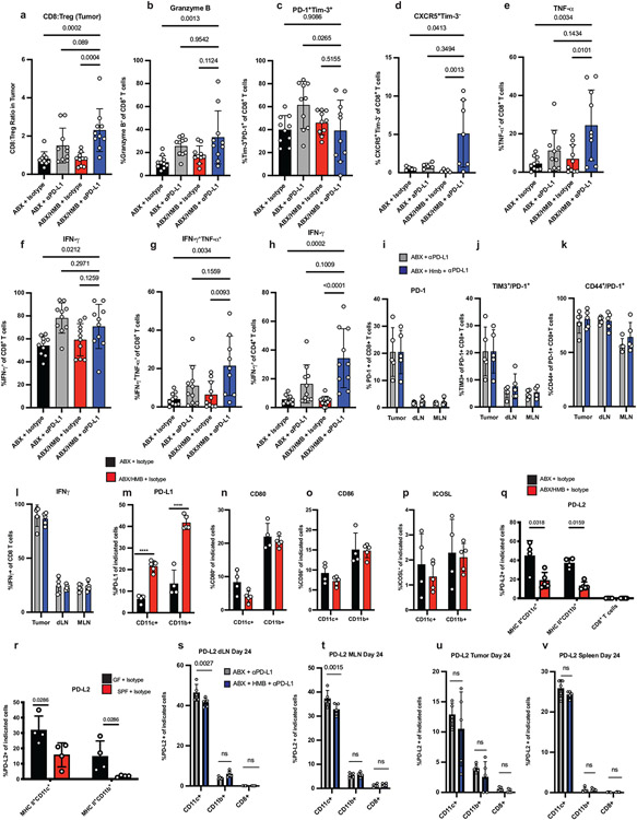
Extended Data Fig. 2. Immune cell responses in ABX, ABX/HMB, GF, and SPF mice.
MC38 tumor cells were implanted subcutaneously in ABX and ABX/HMB mice. Mice were treated with isotype or anti-PD-L1 on days 7, 10, 13, and 16, followed by sacrificed on day 24 after tumor implantation. For detection of cytokines, tumor-infiltrating lymphocytes were stimulated with PMA/Ionomycin for 5 hours in the presence of Golgi inhibitors. (a) Ratio of CD8+ T cells to Treg cells n=10 mice per group (b) Percent of Granzyme B+ CD8+ T cells n= 10 mice per group (c) Frequency of PD-1+TIM-3+ n= 10 mice per group or (d) CXCR5+ TIM-3− among CD8+ T cells in tumors n=5 mice per group for ABX groups and ABX/HMB+ Isotype group and n=7 for ABX/HMB + anti-PD-L1 group. Percent of (e)TNF-α+ n= 10 mice per group (f) IFNγ+n=10 mice per group, and (g) TNF-α+IFNγ+ CD8+ T cells n = 10 mice per group for ABX groups and ABX/HMB + isotype and n= 9 mice for ABX/HMB + anti-PD-L1 (h) Percent of IFNγ+ CD4+ T cells n = 10 mice per group for ABX groups and ABX/HMB + anti-PD-L1 and n=12 mice per group for ABX/HMB + isotype. Mice were analyzed on day 24 after tumor implantation, representative of two independent experiments. Significance measured by non-parametric one-way ANOVA and Dunn’s multiple comparisons and P values are indicated on graphs, error bars show mean and s.d. (i-l) MC38 tumor cells were implanted subcutaneously in ABX and ABX/HMB mice. Mice were treated with isotype or anti-PD-L1 on days 7 and 10 and sacrificed on day 13 after tumor implantation. (i) Percent of PD-1+ CD8+ T cells in tumors, dLNs, and mesenteric lymph nodes (MLNs) n= 5 mice per group (j) Percent TIM-3+ among PD-1+ CD8+ T cells in Tumors, dLNs, and MLNs n= 5 mice per group for all except MLN ABX/HMB + anti-PD-L1 n= 4 mice per group (k) Percent CD44+ expression on PD-1+ CD8+ T cells in tumors, dLNs, and MLNs n = 4 mice per group (l) Percent IFNγ+ CD8+ T cells in tumors, dLNs, and MLNs n= 5 mice per group. (i-l) Significance determined by non-parametric one-way ANOVA with Dunn’s multiple comparisons test, error bars show mean and s.d. Expression of (m) PD-L1, (n) CD80, (o) CD86, and (p) ICOSL on CD11c+ MHCII+ and CD11b+ MHCII+ cells in draining lymph nodes of ABX and ABX/HMB mice implanted with MC38 tumor cells subcutaneously and treated with isotype control mAb as in Figure 1a. Mice were analyzed on day 13 after tumor implantation. N=4 mice in ABX group and n=5 mice in ABX/HMB group. Significance measured by unpaired two-tailed, Mann-Whitney test. Expression of PD-L2 on CD11c+ MHCII+ , CD11b+ MHCII+ cells and CD8+ T cells in draining lymph nodes of (q) ABX vs. ABX/HMB mice n n= 4 mice per group for ABX and n=5 mice per group for ABX/HMB or (r) GF vs. Taconic SPF mice n = 4 mice per group, implanted with MC38 tumor cells subcutaneously and treated with isotype control mAb as in Figure 1a. Mice were analyzed on day 10 after tumor implantation. Significance measured by unpaired, two-tailed Mann-Whitney test, and significant P values indicated on graphs. ABX and ABX/HMB mice were sacrificed 24 days after implantation with MC38 tumor cells. PD-L2 was measured on MHCII+ CD11c+, MHCII+ CD11b+, and CD8+ T-cells in (s) draining lymph nodes n = 5 mice per group, (t) MLN n = 5 mice for ABX and n=4 mice for ABX/HMB, (u) tumors n = 5 mice per group and (v) spleen n= 5 mice per group. Significance measured by Two-Way ANOVA and Sidak’s multiple comparison’s test. Significant P values indicated on graph. Representative experiment of two different experiments. Error bars show mean and s.d.

