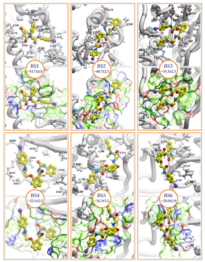
Figure 2.
Binding modes of NCGC607 compound. Poses having the lowest values of binding free energies are shown. Compound and interacting with it amino acid residues are in a balls-and-sticks representation. Hydrogen bonds are depicted by dotted lines. The molecular surface is colored according to binding properties of specific chemical groups within amino acid residues forming the pockets: hydrogen bond donors (blue), hydrogen bond acceptors (red), and hydrophobic groups (green). Circles hold values of NCGC607 binding free energy measured in kcal/mol.

