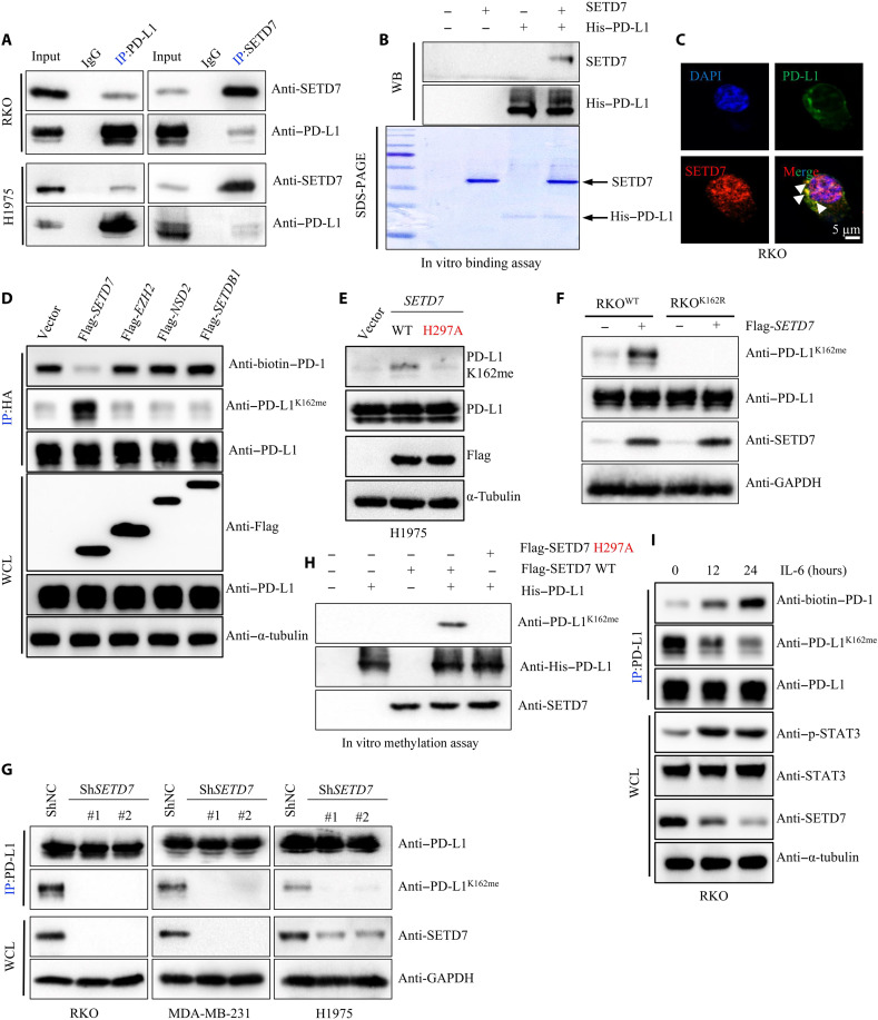
Fig. 3. PD-L1 is methylated at Lys 162 by SETD7.
(A) Whole cell lysis (WCL) were collected for IP with PD-L1 or SETD7 antibody, followed by IB analysis. (B) Coincubating His–PD-L1 and SETD7 proteins, followed by IB analysis. (C) Fixed RKO cells stained with PD-L1 and SETD7 antibodies, followed by immunofluorescence (IF) assays. (D) HEK293T cells transfected with HA–PD-L1, and then cotransfected with Flag-SETD7, Flag-EZH2, Flag-NSD2, or Flag-SETDB1, followed by IP and IB analysis. (E) Transfecting Flag-SETD7 or Flag-SETD7 H297A into H1975 cells, WCE were collected for IB analysis. (F) Transfecting Flag-SETD7 into RKOPD-L1 WT or RKOPD-L1 K162R cells, WCE were collected for IB analysis. (G) Silencing cellular SETD7 expression, WCE were collected for IP with PD-L1 antibody, followed by IB analysis. (H) Immunoprecipitated SETD7 WT or SETD7 H297A protein from HEK293 cells was incubated with S-adenosyl-l-methionine along with His–PD-L1 protein for in vitro methylation assay. PD-L1 methylation was analyzed by IB analysis using with PD-L1 K162 mono-methylation–specific antibody. (I) RKO cells treated by IL-6 (10 ng/ml) in a time-dependent manner, followed by IP and IB analysis. All IBs are performed three times, independently, with similar results.

