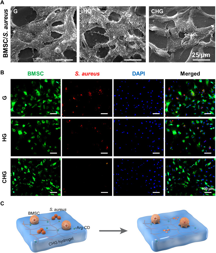
Fig. 5. Competitive colonization assay of cells and bacteria on the surface of composite hydrogels.
(A) Morphology of BMSCs and S. aureus cultured on the surface of composite hydrogels. (B) Images of intracellular bacteria inside the cells, which adhere to the scaffold surface. Green indicates the DiO labeling of BMSCs, red indicates DiD labeling of S. aureus, and blue depicts the 4′,6-diamidino-2-phenylindole (DAPI) staining of the nucleus. (C) Illustration of the cell and bacteria coculture system on the composite hydrogels: CHG composite hydrogel selectively eliminates S. aureus and promotes cell adhesion and growth.

