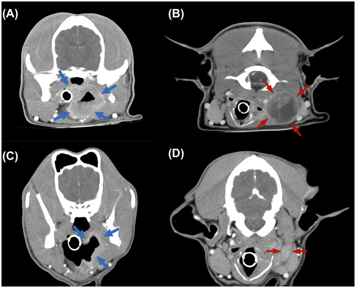
Figure 2.
Transverse computed tomography (CT) images. (A, B) CT images acquired before treatment, revealing the irregular mass from pharynx (blue arrow) and the enlarged medial retropharyngeal lymph node, 3.88 × 3.01 × 3.12 cm in size (red arrow). (C, D) Images from follow-up 4 months CT examination revealing the mass and LN both reached partial remission.

