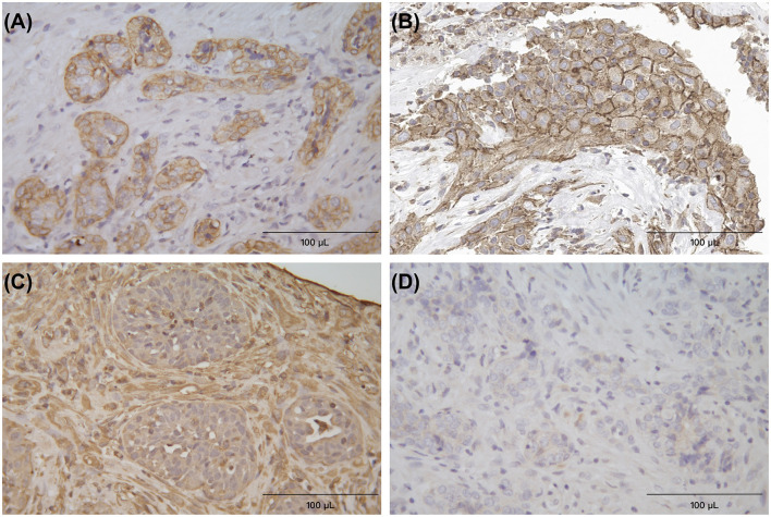
Figure 4.
Immunohistochemical (IHC) staining for cytokeratin (A), PD-L1 (B), vimentin (C), and melan-A (D), respectively, the Horseradish Peroxidase (HRP) method, neoplastic cells formed the glandular appearance and were intensely positive on cytokeratin and PD-L1, and negative on vimentin and melan-A. The surrounding connective tissue cells were positive on vimentin ×400.

