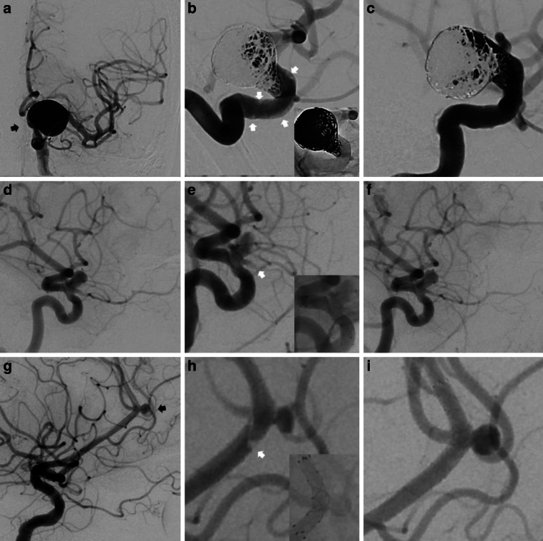
Fig. 1.
Three illustrative cases. a Subtracted AP view of an unruptured aneurysm of the supraophthalmic internal carotid artery (black arrow). b Subtracted and unsubtracted magnified view after coiling and Pipeline Vantage implantation with evidence of in-stent apposition thrombi (white arrows). c Subtracted magnified view after i.v. tirofiban demonstrating complete thrombus dissolvement within the stent. d Subtracted lateral view of a supraophthalmic aneurysm. e Subtracted and unsubtracted magnified view after FRED implantation. Thrombosis of the covered ophthalmic artery (white arrows). f Subtracted magnified view after i.v. tirofiban demonstrating complete thrombus dissolvement. g Subtracted lateral view of an unruptured saccular pericallosal aneurysm (black arrow). h Subtracted and unsubtracted magnified view after FRED Jr. implantation with partially occlusive thrombus visualized at the proximal end in correspondence to the transition zone of the flared ends with the flow diverting segment of the stent (white arrow). i Subtracted magnified view after i.v. tirofiban demonstrates complete thrombus dissolvement

