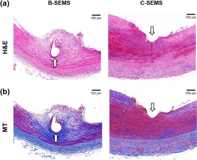
Figure 5.

Representative microscopic images of histologic slices obtained with (a) hematoxylin and eosin and (b) Masson trichrome stains. Degrees of neointimal hyperplasia, inflammatory cell infiltration, and collagen deposition were significantly higher in the C-SEMS group compared to the B-SEMS group. Note Arrow: stent struts, B-SEMS: bare closed-cell self-expandable metallic stent, C-SEMS: covered closed-cell self-expandable metallic stent.
