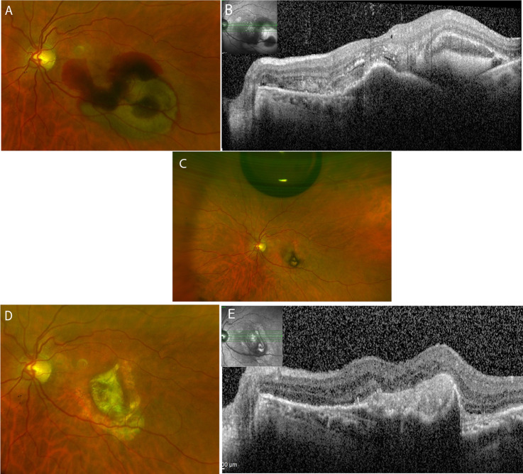
Fig. 1. Colour fundus and OCT over the follow-up of 24 months in a patient intreated with subretinal aflibercept during the surgery.
A Submacular haemorrhage at the day of the surgery. B OCT displaying subretinal haemorrhage and pigment epithelial detachment. C Colour fundus on day 7 after the surgery showing an intravitreal air bubble and resolution of the haemorrhage. D 24 months after the surgery, a macular scar with surrounding atrophy is seen. E The OCT displays the scarring without CNV activity.

