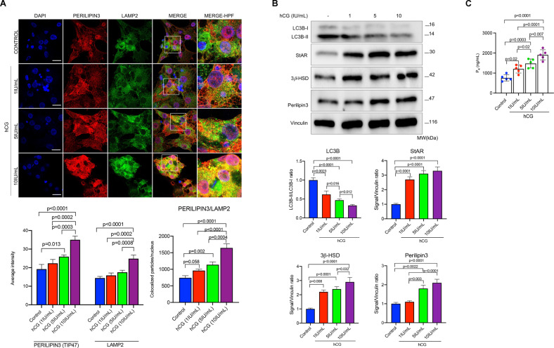
Fig. 4. hCG promotes the association of lipid droplets with lysosomes.
A Representative confocal images of the luteinized granulosa cells 24 h after treatment with hCG at indicated concentrations. Perilipin3 (red signal) and LAMP2 (green signal). Quantification of the signal intensities and co-localizations of the signals are indicated beneath the images. Nuclei stained with DAPI. Scale bars represent 20 μm. Mean ± SD, N = 5 biological replicates analyzed using one-way ANOVA, with Tukey’s test for multiple comparisons. B Representative blots of the luteinized granulosa cells 24 h after treatment with hCG at indicated concentrations. Densitometric quantification is indicated beneath the blots. Mean ± SD, N = 5 biological replicates analyzed using one-way ANOVA, with Tukey’s test for multiple comparisons. C Representative graphic bars indicate progesterone (P4) production of the luteinized granulosa cells treated with hCG at indicated concentrations. Mean ± SD, N = 5 biological replicates analyzed using one-way ANOVA, with Tukey’s test for multiple comparisons.

