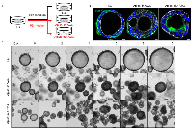
Figure 1.
Generation of apical-out airway organoids. (a) A schematic graph outlines the expansion of lung organoids (LOs) in expansion (Exp) medium and differentiation of apical-in and apical-out airway organoids (AwOs) in proximal differentiation (PD) medium; (b) Bright-field images of the organoids during the expansion or differentiation culture. Scale bar = 200 μm; (c) Confocal images of basolateral marker pan-Keratin (green) in lung organoids and airway organoids. Nuclei and actin filaments were counterstained with DAPI (blue) and Phalloidin-647 (white), respectively. Scale bar = 10 μm.

