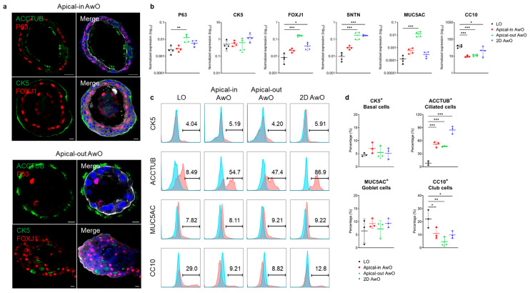
Figure 2.
Characterization of airway organoids. (a) Confocal images of ciliated cell markers (ACCTUB, green; FOXJ1, red) and basal cell markers (P63, red; CK5, green) in apical-in (top) and apical-out (bottom) airway organoids (AwOs). Nuclei and actin filaments were counterstained with DAPI (blue) and Phalloidin-647 (white), respectively. Scale bar = 10 μm. (b) Non-differentiated lung organoids (LOs) and differentiated airway organoids (AwOs) were assessed through qPCR analysis to detect the expression level of marker genes specific for basal (P63, CK5), ciliated (FOXJ1, SNTN), goblet (MUC5AC) and club (CC10) cells. Data represent means ± SD of a representative experiment, n = 4. Ordinary one-way ANOVA with Dunnett’s multiple comparison test comparing airway organoids to the lung organoids. * p ≤ 0.05, ** p ≤ 0.01, *** p ≤ 0.001. (c,d) Non-differentiated lung organoids (LOs) and differentiated airway organoids (AwOs) were assessed through flow cytometry to examine the abundance of CK5+ basal cells, ACCTUB+ ciliated cells, MUC5AC+ goblet cells and CC10+ club cells. Representative histograms are shown in (c): red, cells stained with target antibodies; blue, cells stained with isotype controls. Data shown in (d) represent means ± SD of a representative experiment in one organoid line, n = 3. Ordinary one-way ANOVA with Dunnett’s multiple comparison test comparing airway organoids to the lung organoids.

