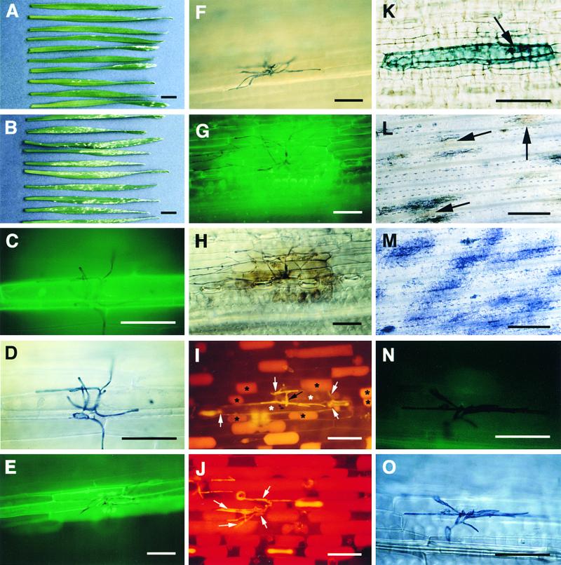
Figure 3.
Effect of Syringolin A on the Wheat–Powdery Mildew Interaction.
(A) and (B) Leaves of powdery mildew–inoculated plants treated with 40 μM syringolin A (A) or a control solution (B) at 2 days after inoculation. The photograph was taken 7 days after inoculation.
(C) to (H) Autofluorescence at infection sites on leaves treated with syringolin A at 2 days ([C] and [E]) or 3 days (G) after inoculation. The same infection sites under bright-field illumination are shown in (D), (F), and (H), respectively.
(I) and (J) Infection sites on leaves treated with 40 μM syringolin A (I) or a control solution (J) at 2 days after inoculation. Leaves were infiltrated with a hypertonic solution containing neutral red dye at 5 days (I) and 1 day (J) later, when fungal development was approximately equal. Live cells plasmolyze under these conditions and take up neutral red dye. The black arrow in (I) points to fungal structures (appearing yellow); black asterisks mark the protoplasts of live cells surrounding two dead cells, which are marked with white asterisks. The white arrows indicate the left and right borders of the two dead cells, which do not contain a protoplast. The white arrows in (J) point to fungal structures. All cells at the infection sites are plasmolyzed and stained red.
(K) Infected dead cell on a leaf treated with syringolin A at 2 days after inoculation and stained with trypan blue 5 days later. The arrow points to the haustorium. No other fungal structures are visible.
(L) and (M) Infections sites on a plant treated with syringolin A (L) or a control solution (M) at 4 days after inoculation. Leaves were photographed under bright-field illumination. The arrows in (L) point to examples of infection sites where tissue browning is visible.
(N) and (O) Infection site on a plant treated with 200 ppm (888 μM) cyprodinil and photographed under incident fluorescent light (N) or in the bright field (O). The image in (N) has been overexposed because of the absence of significant autofluorescence.
In (C) to (H) and (L) to (O), the leaves shown were stained with lactophenol/trypan blue for 1 hr before photography to stain fungal structures. In (A) to (I) and (K) to (O), the photographs shown were taken 7 days after inoculation.  ;
;  ;
;  .
.

