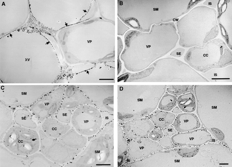
Figure 8.
Cytochemical Localization of Wound-Inducible H2O2 in Vascular Bundles of Tomato Leaves.
(A) Electron-dense deposits of CeCl3 indicative of the presence of H2O2 in developing secondary cell walls of xylem vessels (XV) of control unwounded leaves (arrows show typical deposits). Note that the cell walls of an adjacent vascular parenchyma cell (VP) show little CeCl3 staining.
(B) Absence of CeCl3 staining in the cell walls of vascular parenchyma (VP) and neighboring spongy mesophyll (SM) cells associated with the phloem in control unwounded leaves.
(C) H2O2 generation in the vascular bundle of a wounded tomato leaf 4 hr after wounding. H2O2 accumulates strongly in the cell walls of vascular parenchyma cells bordering spongy mesophyll cells and at the intercellular spaces (IS).
(D) Systemic accumulation of H2O2 in vascular bundles of upper unwounded leaves of young tomato plants 4 hr after wounding of the lower leaf.
CC, companion cell; CW, cell wall; SE, sieve element.  .
.

