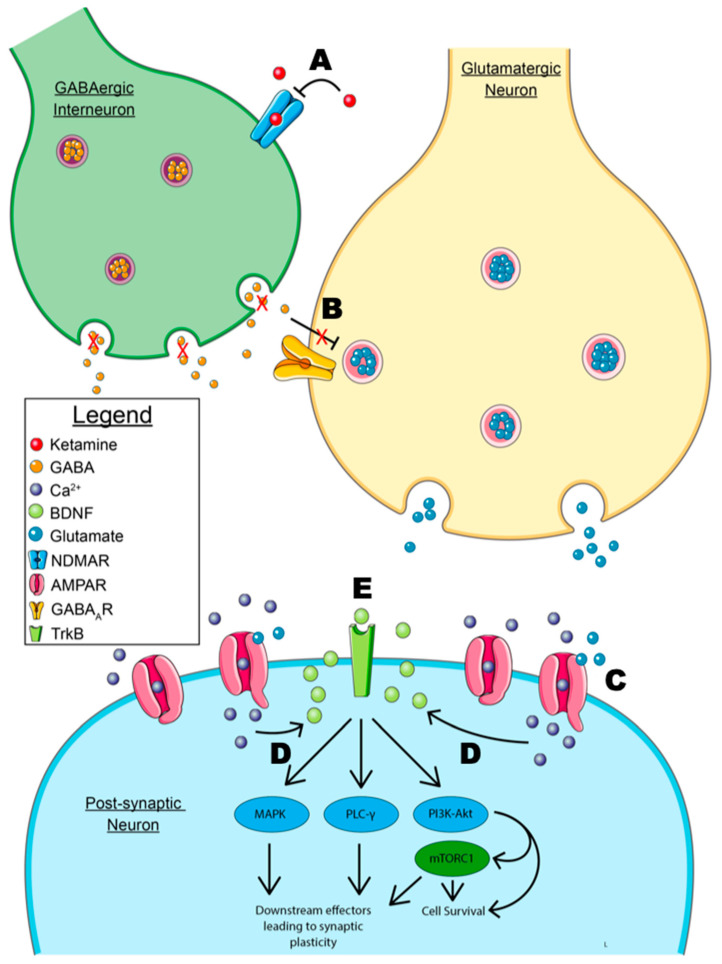
Figure 2.
Model of the disinhibition hypothesis. A. Ketamine’s antidepressant mechanism of action primarily depends on the antagonism of NMDARs (N-methyl-D-aspartate receptors) on GABAergic interneurons preventing GABA release. B. The inhibition of GABA release prevents the inhibition of pyramidal glutamatergic neurons. This allows for the release of glutamate and the downstream effects of the subsequent glutamate surge. C. Glutamate binds to post-synaptic AMPARs (α-amino-3-hydroxy-5-methyl-4-isoxazolepropionic acid receptor), allowing for calcium influx. D. Calcium influx leads to the calcium-dependent release of BDNF from the post-synaptic membrane. NOTE: the model shows the post-synaptic release of BDNF, though an immunofluorescent localization study has suggested that the pre-synaptic release of BDNF at the downstream synaptic cleft is involved [112]. E. Autocrine signaling of BDNF leads to downstream signaling through the MAPK, PLC-γ, and PI3K-Akt signaling pathways. The MAPK and PLC-γ pathway are primarily implicated in synaptic plasticity, while the PI3k-Akt pathway leads to anti-apoptotic signaling and cell survival. Signaling through mTORC1 has also been implicated in synaptic plasticity and neuritogenesis. NOTE: As mentioned previously, autocrine signaling is shown, but paracrine signaling may be involved based on the location of BNDF-containing vesicles in immunofluorescence studies [112]. Some graphic components from Servier Medical Art were used to draw parts of this model. Servier Medical Art by Servier is licensed under a Creative Commons Attribution 3.0 Unported License (https://creativecommons.org/licenses/by/3.0/, accessed 3 May 2023).

