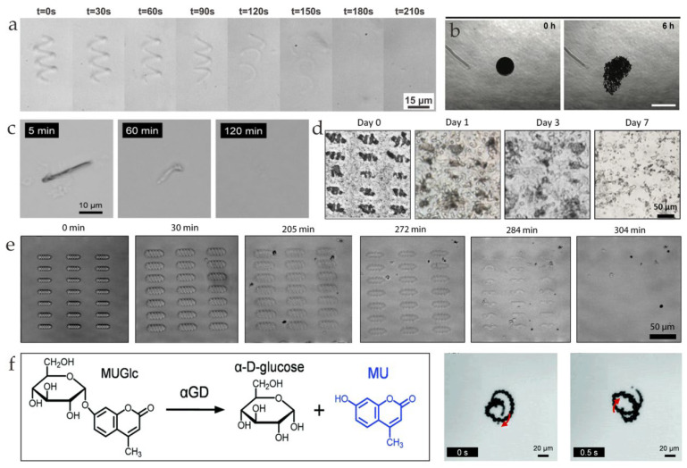
Figure 5.
(a) Degradation of a GelMA helical microstructure in a collagenase solution (0.1 mg mL−1) (reprinted with permission from Ref. [29]). (b) Enzymatic biodegradation and magnetic retrieval of SPIONs from the GelMA microrobot in the absence of hNTSCs (reprinted with permission from Ref. [75]). (c) Transformation of the morphology of Avi/bUre microtube in pronase solution at 37 °C. (reprinted with permission from Ref. [77]). (d) Optical images displaying the degradation process of magnetoelectric (ME) soft helical microswimmers after being cultured with cells for 0, 1, 3, and 7 days (reprinted with permission from Ref. [76]). (e) Differential interference contrast (DIC) images of a degrading microswimmer array with 4 μg/mL of enzyme (reprinted with permission from Ref. [30]) (f) Microscopic observations of self-propulsion of aGD/Cat MTs through the jetting of O2 bubbles in phosphate buffer (PB) solution and hydrolysis reaction of MTs. The red arrows indicate the direction of O2 bubbles (reprinted with permission from Ref. [78]).

