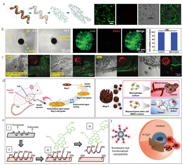
Figure 8.
(a) Illustration of microswimmer’s degradation process and induced neuronal differentiation of SH-SY5Y cells (reprinted with permission from Ref. [76]). (b) Live/dead cell imaging of the hNTSCs on the microrobot (left), images after incubating cells with microswimmers (middle), and evaluation of cell viability (right) (reprinted with permission from Ref. [75]). (c) Confocal scans of histological sections of skin tissues implanted with degradable 75 vol% polyethylene glycol diacrylate (PEGDA):25 vol% pentaerythritol triacrylate (PETA) microrobot and hard-to-degrade 100 vol% PETA microrobot. The red arrows indicate the location of the microrbot (reprinted with permission from Ref. [73]). (d) Preparation and in vivo locomotion of the radiocontrast-agent-loaded magnetic chitosan microscaffold (Mag-C) for real-time X-ray imaging (left) and schematics of Mag-C containing macrophages and human adipose-derived mesenchymal stem cells (hADMSCs) used for cancer therapy and cartilage regeneration (right) (reprinted with permission from Ref. [101]). (e) Conjugation of NIR−797 dyes to ABFs for functionalization (reprinted with permission from Ref. [102]). (f) Schematic illustration of fluorescent-dye-coated magnetic nanoparticles and the generation and navigation of swarm inside cell. The black arrows indicate the direction of the magnetic field (reprinted with permission from Ref. [103]).

