Figure 2.
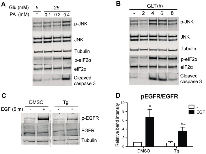
ER stress impairs EGFR phosphorylation. (A) To verify induction of ER stress, 832/13 cells were cultured in media containing 5 mM glucose and BSA or 25 mM glucose and increasing concentrations of palmitic acid complexed to BSA (0, 0.1, 0.2, or 0.4 mM) for 8 h. (B) In a complementary experiment, cells were exposed to 25 mM glucose and 0.4 mM palmitic acid for up to 8 h. Cells were harvested, and lysates were immunoblotted using antibodies directed against p-JNK, JNK, p-eIF2α, eIF2α, and cleaved caspase 3 to establish the extent of ER stress produced by glucolipotoxicity. Shown are representative, confirmatory immunoblots. (C) To induce ER stress, 832/13 cells were treated with DMSO (vehicle control) or 1 μM thapsigargin (Tg) for 4 h, followed by starvation and EGF stimulation as before. Protein levels of p-EGFR, EGFR, and tubulin were analyzed by immunoblotting. Representative blots of n ≥ 3 experiments are shown, and results are quantified in (D). Groups were compared using ANOVA with Bonferroni post hoc tests. * p < 0.05 vs. BSA, non-stimulated; # p < 0.05 vs. BSA, EGF-stimulated.).

