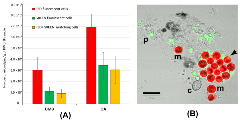
Figure 3.
(A) Graphs of the number of microalgae (zooxanthellae) measured in the tissues of umbrellas (UMB) and oral arms (OA) of C. andromeda. (B) Confocal microscope image of the lyophilized jellyfish tissue resuspended in seawater. Red-autofluorescent chloroplasts in microalgae (m), green-autofluorescent particles (p), and a cnidocyst (c) are visible. The autofluorescent particles are automatically counted as red, green, and red + green autofluorescence. The round-shaped red-autofluorescent cells with a diameter around 10 μm were considered as microalgal cells. Bar = 20 μm.

