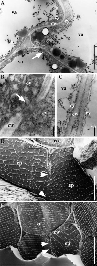
Figure 3.
prc1 Dark-Grown Hypocotyls Display Abnormal Cell Wall Structures.
(A) to (C) Transmission electron micrographs of cross-sections from dark-grown hypocotyls (4 days old) of prc1-1 mutant ([A] and [B]) and wild-type (C) seedlings in the cortical cell layer. Arrow in (A) indicates an abnormal wall portion shown at a higher magnification in (B).
(D) and (E) Scanning electron micrographs of prc1-1 dark-grown hypocotyls. Arrowheads indicate protruding stubs of incomplete cell walls in the epidermal cell layer.
co, cortex cell; cw, cell wall; cy, cytoplasm; ep, epidermal cell; va, vacuole.  ;
;  ;
;  ;
;  .
.

