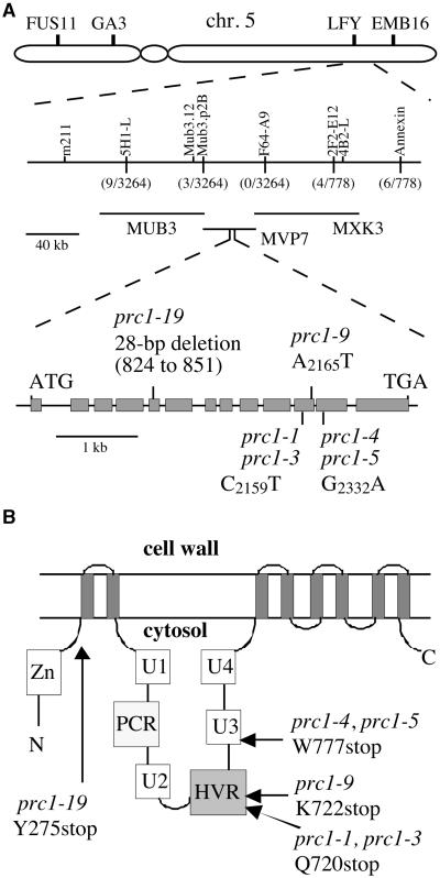
Figure 6.
The PRC1 Gene Is Isolated Through Map-Based Cloning.
(A) Physical map of the region of chromosome (chr.) 5 containing the PRC1 gene. The prc1-1 mutation was mapped to the bottom of chromosome 5, south of molecular marker LFY. New markers positioned the prc1-1 mutation between markers Mub3.p2B and 2F2-E12. The number of recombinant chromosomes is indicated in parentheses. Sequence analysis of candidate genes between these two markers showed that six prc1 mutant alleles carried a point mutation or a deletion in the CesA6 gene carried by P1 clone MVP7. The PRC1 (CesA6) gene comprises 13 exons (filled boxes) and 12 introns. The prc1 mutations that were identified are indicated.
(B) Predicted topology of the PRC1 protein. The protein is predicted to contain eight membrane-spanning helices (indicated by the eight gray bars). The putative Zn binding domain, the U1, U2, U3, and U4 domains, the plant conserved region (PCR), and the HVR are predicted to face the cytosol (Delmer, 1999). The positions of the mutations are indicated. N, N terminus; C, C terminus.

