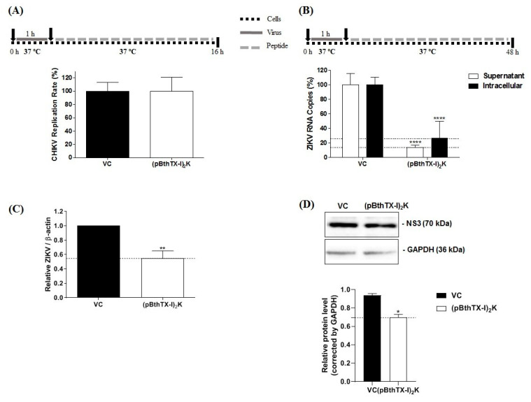
Figure 4.
Analysis of the effect of the (p-BthTX-I)2K peptide on the post-entry stages of CHIKV and ZIKV infection. (A) Effect of peptide treatment at 12.5 µM on the post-entry steps of CHIKV-NLuc in BHK-21 cells. (B) Effect of the peptide treatment at 25 µM on the post-entry stages of ZIKV in Vero cells as measured by the amount of virus RNA in the supernatant and infected cells. (C) Normalization of ZIKV RNA levels on the basis of the ratio of the total intracellular RNA to the mRNA of β-actin. CHIKV replication was analyzed via the measurement of NLuc activity 16 h.p.i. The levels of ZIKVBR RNA were quantified via qRT–PCR 48 h.p.i. (D) NS3 ZIKV protein expression in the post-entry assay. The level of this protein was quantified using Western blot analysis. Error bars represent ± SD. The mean values ± SDs represent data from a minimum of three independent experiments that were each performed in quadruplicate for the CHIKV assay and in duplicate for the ZIKV assay. * p ≤ 0.05; ** p ≤ 0.01; **** p ≤ 0.0001. VC: vehicle control (sterile water). A schematic representation of the respective experiment is shown above each graph.

