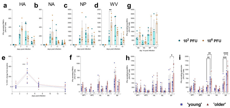
Figure 4.
Cellular response to H1N1 GM19 influenza infection in the ferret and Golden Syrian hamster. PBMCs were collected at baseline and days 7, 11 and 14 post-infection to assess longitudinal IFNγ responses. PBMCs were stimulated with (a) HA, (b) NA, (c) NP and (d) whole virus (GM19). A significant (** p = 0.0052) difference between the 106 PFU (high)-dose-infected ferrets and the 102 PFU (low)-dose-infected ferrets was detected at day 7 post-infection when PBMCs were stimulated with homologous virus. Statistical analysis was performed used mixed-effects analysis. (e) Small blood volumes were collected from the gingiva of hamsters at baseline and days 1, 4, 7 and 14 post-infection. The percentage change of CD4+ cells from baseline was calculated, and a significant (**** p < 0.0001) increase in cells was observed in both ages of hamsters. Statistical analysis was performed using mixed-effects analysis. Symbols show individual hamsters; lines show means. (f) PBMCs were collected at cull to assess IFNγ responses. PBMCs were isolated from hamsters at days 4 and 14 post-infection and were stimulated with M1, M2, HA, NA, and NP. (g) At 14 days post-infection, ferret lung MNCs were assessed for influenza-specific IFNγ responses. Bars show mean; error bars show SD. (h) Hamster lung MNCs * p < 0.05 and (i) splenocytes were isolated at days 4 and 14 post-infection and were stimulated with M1, M2, HA, NA, and NP. A significant increase from day 4 to 14 in HA- (** p < 0.01) and NP- (*** p = 0.0005, **** p < 0.0001) specific IFNγ responses in both young and older hamsters, respectively, was observed in splenocytes. A significant increase from day 4 to 14 in NP- (* p = 0.0112) specific IFNγ responses in older hamsters was also observed in lung MNCs. Statistical analyses were performed using two-way ANOVA. Bars show mean; error bars show SD.

