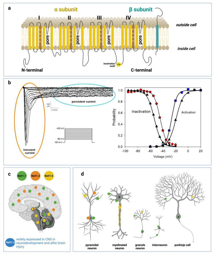
Figure 1.
Voltage-gated sodium channel structure, function and distribution in CNS: (a) Schematic representations of α-subunit and auxiliary β-subunit of Nav channels. The α-subunit of the channel consists of four homologous domains (DI–DIV) made of six transmembrane helices (S1–S6). The voltage-sensor is localized in the fourth helix (S4) in each domain. The loops between S5 and S6 in each domain form the pore. (b) Left side: representative current traces recorded from a HEK293 cell transiently transfected with a cDNA encoding Q1489H/Nav1.1 mutant channel. Current traces were evoked from a series of 20 ms depolarizing pulses from −80 to +50 mV in 10 mV increments, starting from a holding potential of −90 mV. Q1489H mutation exhibits a large persistent current. Right side: curves of voltage dependence of steady-state activation (blu squares) or inactivation (red circles) are represented. The same curves are represented also for a WT-SCN1A transfected cell (black triangles) not reported in the figure. Lines stand for the Boltzmann function fits (c) Distribution of Nav1.1, Nav1.2, Nav1.3, and Nav1.6 in human brain regions. (d) Cellular and subcellular distribution of Nav1.1, Nav1.2, Nav1.3, and Nav1.6 in human brain.

