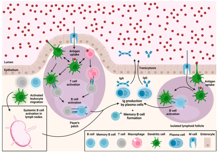
Figure 1.
Antibody response to mucosal antigens. Antigens (shown above as red spheres) are sampled in the GALT by DCs and M cells in Peyer’s patch (left) and ILFs (right). Peyer’s patches heavily use T cells to activate B cells, whereas ILFs do not require T cells. B cells can also be activated in lymph nodes (bottom left) following the migration of activated B cells, T cells, and/or DCs from the GALT. The above diagram is not to scale and does not show all mucosal immune processes. A publication license for BioRender content used in the above figure was obtained on 24 February 2023 (agreement number NO251WZ00A).

