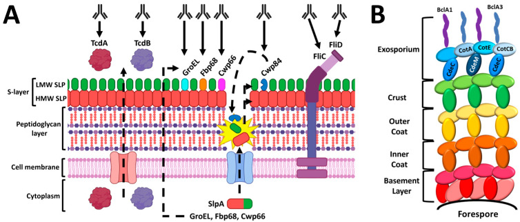
Figure 3.
C. difficile vaccine candidates. Diagram of toxin and cell-surface antigens (A) and spore coat proteins (B) currently under investigation. The structure of the C. difficile cell envelope shown here (A) is based, in part, on the illustrations provided by a prior review [109]. Dotted lines indicate generalized transport mechanisms that are not completely understood. The S-layer is composed of the high molecular weight S-layer protein (HMW SLP) and an outer layer of low molecular weight SLPs (LMW SLPs) and additional proteins [110]. The CD0873 lipoprotein, despite being a mucosal vaccine candidate, is not shown because its precise position in the S-layer of C. difficile has not yet been determined [109]. All spore-coat vaccine candidates (B) are localized in the exosporium layer. The spore structure diagram (B) was based on a recent review by Paredes-Sabja et al. [111]. A publication license was obtained for BioRender content used in Figure 3A on 2 January 2023 (agreement number LX24UCVM8E).

