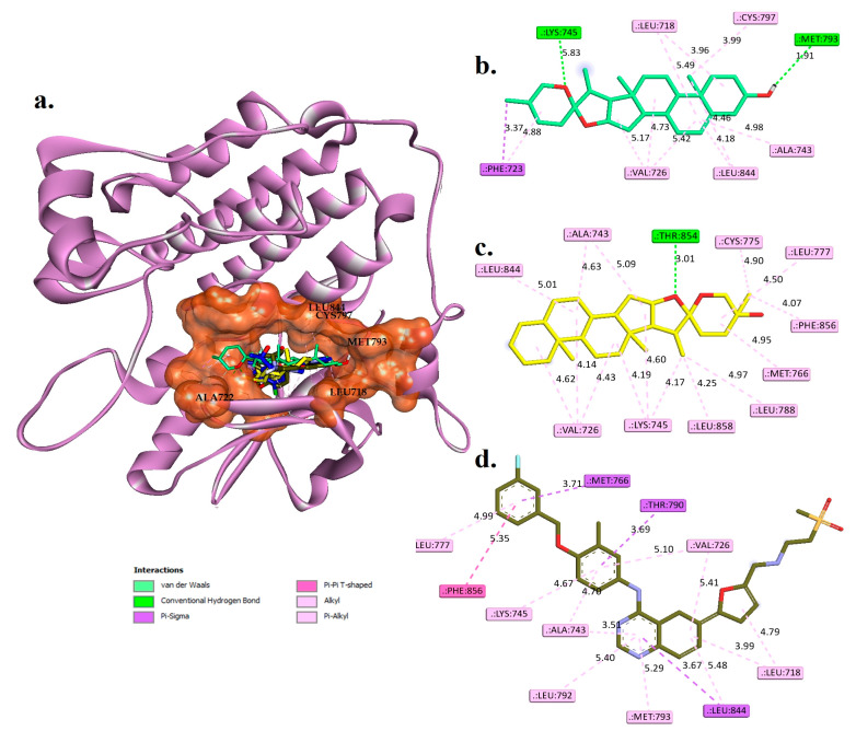
Figure 3.
(a) The docked structures of diosgenin (coloured green C and red O), monohydroxy spirostanol (coloured yellow C and red O), lapatinib (coloured army green C, red O, stune N, orange S, and cyan F), and the original co-crystallized tak-285 (coloured dark blue C, red O, stune N, green CL, and cyan F) are superimposed into the active binding site (coloured rust) of EGFR (3POZ.PDB). (b–d) Two-dimensional interaction views of the key and surrounding amino acids with diosgenin, monohydroxy spirostanol, and lapatinib, respectively.

