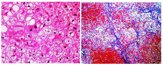
Figure 1.
Liver histology of CD patient. Hematoxylin and eosin staining (left, ×100) revealed microvesicular and macrovesicular steatosis and ballooned hepatocytes. Azan–Mallory staining (right, ×40) showed marked pericellular fibrosis with disorganization of hepatic lobules. This figure is reprinted/adapted with permission from Ref. [24]. 2007 by the AGA Institute.

