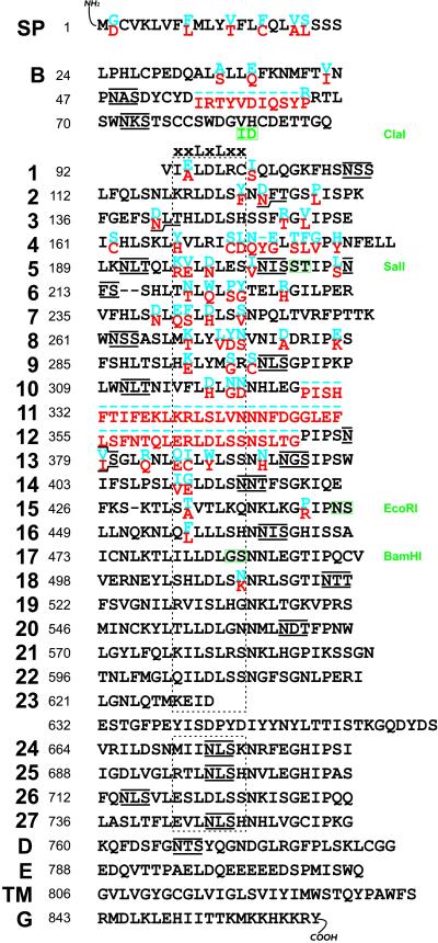
Figure 1.
Primary Structure and Alignment of Cf-4 and Cf-9 Resistance Proteins.
Amino acid residues of Cf-4 and Cf-9 that are identical are shown in black. Cf-4– and Cf-9–specific residues are shown in blue and red, respectively. The numbering (left) corresponds to the Cf-9 protein sequence. Potential N-glycosylation sites (NxS/T) in Cf-4 and Cf-9 are overlined and underlined, respectively. The stippled box indicates the various β-sheets (consensus xxLxLxx), each of which contains five solvent-exposed amino acid residues (x). Residues encoded at restriction sites are indicated with green boxes. Introduction of a ClaI site resulted in two residue changes, whereas the SalI and BamHI sites were introduced as silent mutations. The EcoRI site is endogenous for Cf-4 and Cf-9. Domains, indicated at left, are as follows: SP, signal peptide (A-domain); B, cysteine-rich domain; 1 to 27, LRRs; D, domain without conspicuous features; E, acidic domain; TM, putative transmembrane domain; G, basic domain, representing the putative cytoplasmic tail.

